'Designed A Special Kind Of Incision': Doctors Remove 1.9-Kg Tumour From 17-Year-Old’s Chest In Gurgaon Hospital
A high-resolution computed tomography (CT) scan revealed that a rare form of tumour was occupying two-thirds of the chest cavity of a 17-year-old boy. The tumour is called a thymolipoma.

In a breakthrough in the field of medical science, doctors at the Fortis Memorial Research Institute (FMRI) in Gurugram have successfully removed a 1.9-kilogram tumour from the chest cavity of a 17-year-old boy. The patient, Saksham Chauhan, complained of neck and chest pain, and suffered from fever, following which the doctors at Fortis Gurugram conducted several medical tests. Initially, they suspected that Chauhan was suffering from typhoid fever, a disease characterised by fever, headache, and stomach pain.
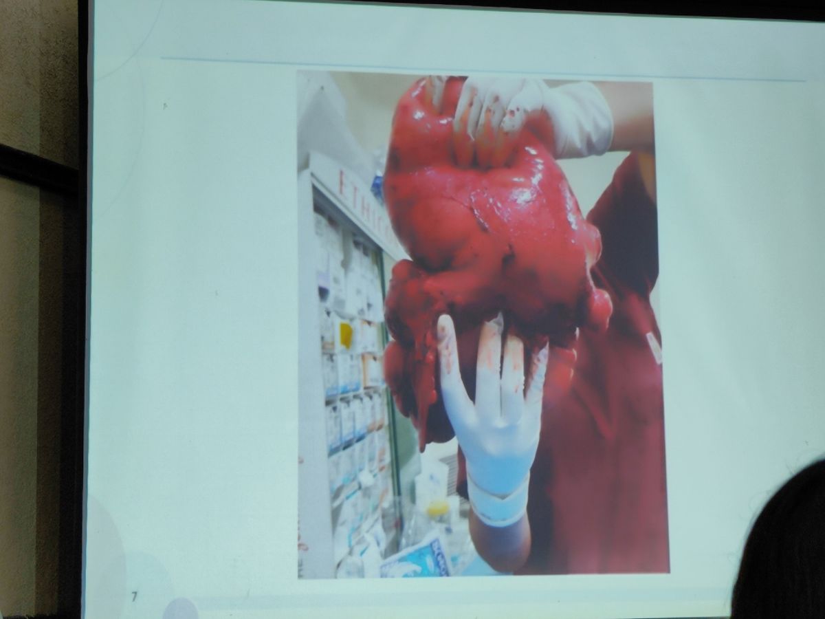
However, a high-resolution computed tomography (CT) scan revealed that a rare form of tumour was occupying two-thirds of his chest cavity. The tumour is called a thymolipoma.
What is a thymolipoma?
A thymolipoma is a rare, benign mass of thymic origin, and contains both thymic and mature adipose tissue. It is formed in the anterior portion of the mediastinum, or thoracic cavity. A thymolipoma was formed in the thoracic cavity of Chauhan because his thymus gland grew in size, and covered huge portions of the chest and lungs. It is believed that due to a growth hormone imbalance, lymphocytes had started growing at a fast pace, causing the tumour to be formed.
What problems did the patient face due to the tumour?
Since the tumour applied a large amount of pressure on the lungs and the heart, the organs were not functioning properly.
Moreover, Chauhan weighed less than 30 kilograms.
What made the surgery more complex than similar instances of tumour removal?
Led by Dr Udgeath Dhir, Director and Head of Cardiothoracic and Vascular Surgery, Fortis Gurugram, the doctors performed a complex surgery and successfully removed the thymolipoma from the chest cavity of the boy.
On the sidelines of a press conference held to brief people about the breakthrough, ABP Live spoke to Dr Dhir, and Dr Anand Kumar, Director, Cardiac Anaesthesia, Fortis Gurugram, and asked them how complex the surgery was compared to similar cases of tumour removal.
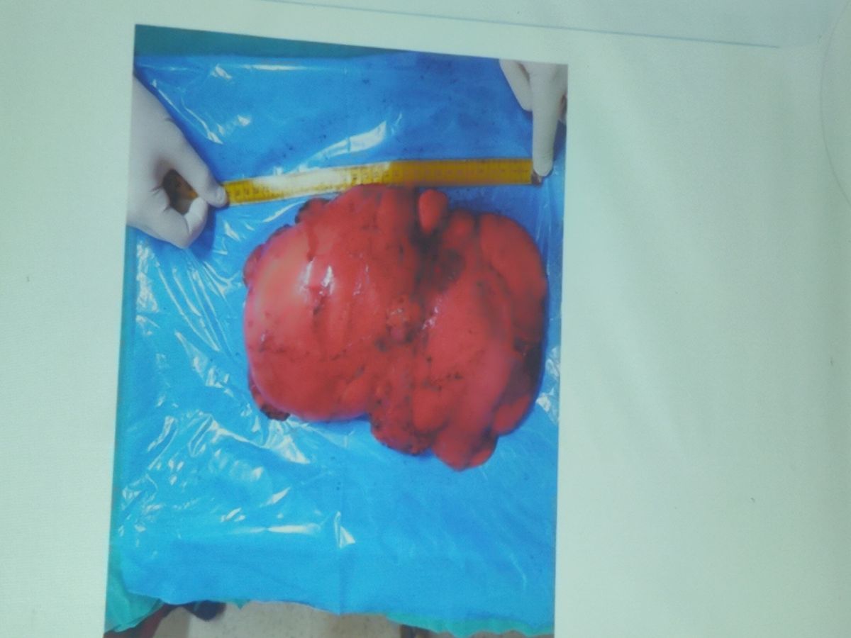
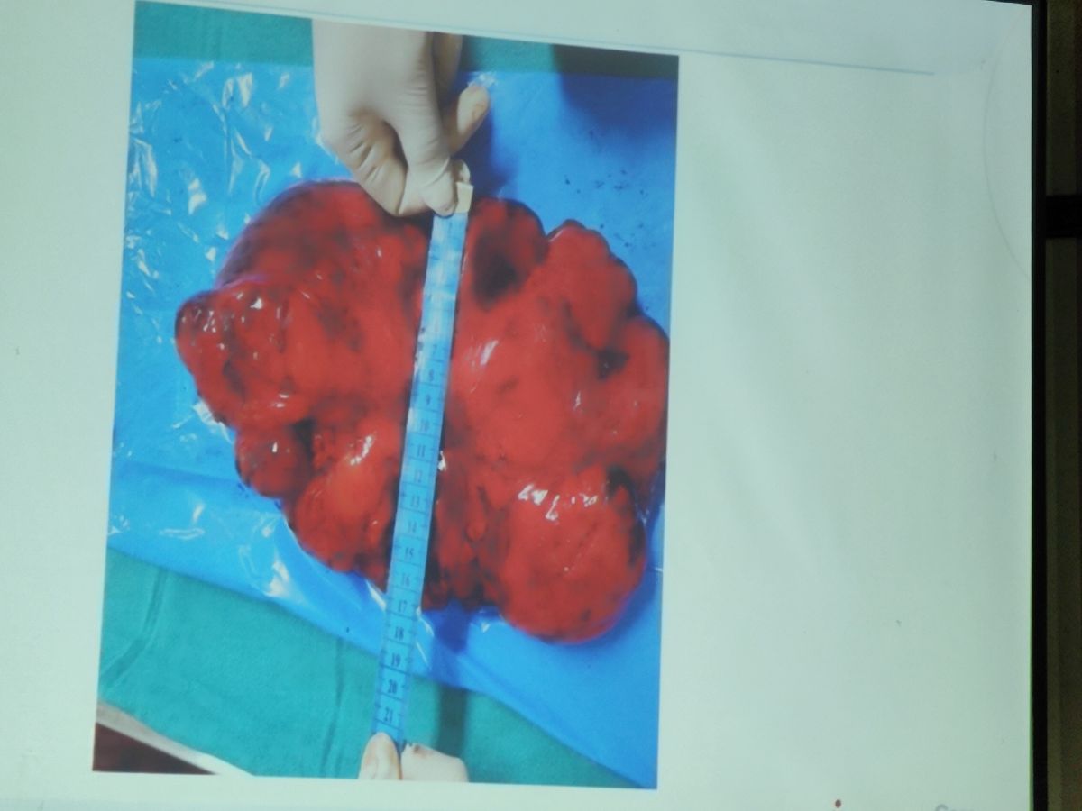
Dr Dhir said that the tumour, whose dimensions were 30 cm × 18 cm × 8 cm, had occupied two-thirds of the chest cavity, and was attached to the lower end of the space. It was because of this that the team of doctors had to "design a special kind of incision" to remove the tumour. "Since a large tumour was occupying two-thirds of the chest cavity and was attached to the lower end of the cavity, it was not possible for us to approach the tumour through a minimally invasive surgery. Hence, we designed a special kind of incision called clamshell incision, and we approached the tumour through that."
Clamshell incision, also known as transverse or crossbow trans-sternal incision, is a surgical technique used to approach the heart or to access mediastinal (thoracic cavity) tumours or both the lungs. This technique was commonly used in the early days of cardiac surgery to reach the heart, according to the US National Institutes of Health (NIH). The doctors at Fortis made incisions along the sides of the lungs to access the tumour.
Explaining what made the surgery more complex compared to other instances of tumour removal, Dr Dhir said, "First, we have to remove the tumour from the chest wall it was adhered to. The tumour was compressing almost two-thirds of the lungs. The heart was completely compressed. So, such tumours misbehave and any mishap could have been extremely detrimental. This signifies the complexity of the surgery."
MUST READ | Nipah Virus: How Fast The Bangladesh Variant Spreads And Who Is At Greatest Risk? Know Symptoms, Treatment
Another challenging aspect of the surgery was the administration of anaesthesia. Anaesthesia had to be administered in such a way that the heart would not stop pumping blood. It was due to the huge mass of the tumour that anaesthesia administration proved to be so challenging.
Explaining how this challenge was overcome, Dr Kumar said, "We had to monitor him by placing some invasive lines. At every moment, we monitored his heartbeat, blood pressure, and oxygen levels. Anaesthesia was given in a careful manner, and he did not feel any pain, neither intra-operatively nor post-operatively."
MUST READ | The Science Of Health: How Genetics And Mutations Determine One's Susceptibility To Cancer
It took the doctors 3.5 hours to perform the surgery.
If anaesthesia were not administered carefully, the heart and respiratory muscles would have been paralysed, and blood circulation would have stopped. Dr Dhir said that the respiratory muscles were taken care of during the administration of anaesthesia.
Are there any chances of the tumour reappearing?
Dr Dhir said that every cell of the tumour, which is likely to have formed over a period of 1.5 to two years, has been removed, and the chance of recurrence is less than 0.001 per cent.
What precautions has the patient been advised to take?
Chauhan was discharged from the hospital five days after the successful surgery. Dr Dhir said that some of the post-surgery precautions the patient has been asked to take include avoiding heavyweight lifting and stretching. He has also been asked to undergo lung physiotherapy.
Check out below Health Tools-
Calculate Your Body Mass Index ( BMI )
Trending News
Top Headlines


